
Histopathology & Cytology in Lucknow
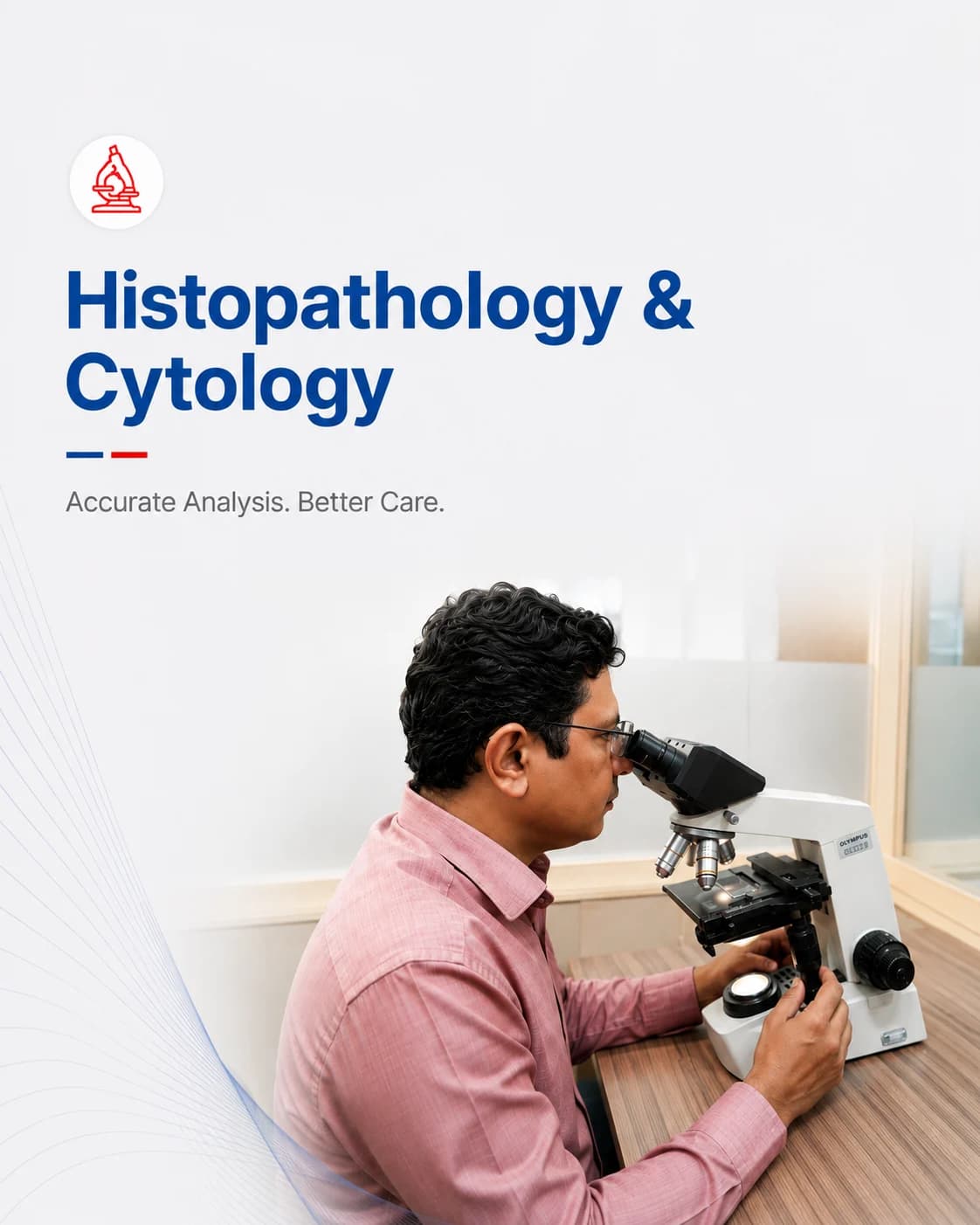
Examining tissue and cells under the microscope to reach a definite diagnosis.
Histopathology is the examination of tissue under the microscope, and cytology the examination of individual cells. When a doctor needs certainty — is this lump benign or not, what exactly is this growth — this is the test that gives the definitive answer.
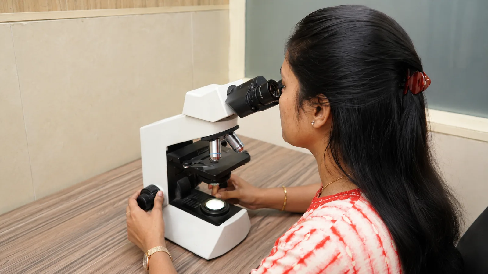
At Charak, biopsy and cytology specimens are processed carefully and reported by consultant pathologists. This is the most human of laboratory work: the diagnosis is made by a trained eye reading the tissue, and difficult cases are reviewed before the report is signed.
Histopathology, biopsy and cytology explained
A biopsy takes a small piece of tissue, which is thinly sliced, stained and examined to diagnose the nature of a growth or disease — it is how cancers are confirmed and graded, and how many skin, gut and organ conditions are named.
Cytology examines loose cells instead of solid tissue: a Pap smear screens the cervix, an FNAC samples a lump with a fine needle, and fluid cytology examines cells in a body fluid. Less invasive, and often the first step.
Your doctor may order these for
- A biopsy of a lump, ulcer, skin lesion or internal growth
- FNAC of a thyroid nodule, breast lump or enlarged lymph node
- A Pap smear for cervical cancer screening
- Examining fluid drawn from the chest or abdomen
- Confirming and grading a suspected cancer
How to prepare
The tissue or sample is usually taken by your treating doctor or during a procedure; the laboratory then processes and reports it. For a Pap smear, timing and simple preparation improve the sample.
- Pap smear: avoid the days of your period, and do not use vaginal medication for two days before
- For an FNAC done at the centre, no special preparation is usually needed
- Bring the clinical details and any imaging — they guide how the tissue is interpreted
- Bring previous biopsy reports or slides if you have them, for comparison or review
What to expect
- 01
Sample received
The biopsy, smear or fluid arrives at the lab, fixed and labelled with your clinical details.
- 02
Processing
Tissue is processed, thinly sectioned and stained; smears are fixed and stained for the microscope.
- 03
Microscopic examination
A consultant pathologist examines the slides and, for difficult cases, seeks a second opinion before reporting.
- 04
Detailed report
A descriptive report with the diagnosis is issued — the turnaround is longer than blood tests because the work is meticulous.
Common questions
Why does a biopsy report take several days?
Tissue must be fixed, processed, sectioned and stained before a pathologist can examine it — each step takes time and cannot be shortened without compromising the diagnosis. Difficult cases are also reviewed before signing.
What is the difference between a biopsy and an FNAC?
A biopsy removes a small piece of solid tissue; an FNAC uses a fine needle to draw out cells from a lump. FNAC is quicker and less invasive, and is often the first step before a full biopsy if needed.
How should I prepare for a Pap smear?
Book outside your period, and avoid vaginal medication or douching for two days beforehand, as these can affect the cells collected and the quality of the result.
From the health library
Plain-language guides written by the team here — what the test measures, how to prepare, and what the result means.
Related services
Microbiology
Finding the organism behind an infection — and the antibiotic that will treat it.
Read more →
Serology & Immunology
Antibody and antigen tests for infections, immunity and autoimmune conditions.
Read more →
Molecular Diagnostics
Detecting infections and genetic changes at the level of DNA and RNA.
Read more →