
Gamma Camera & SPECT Imaging in Lucknow
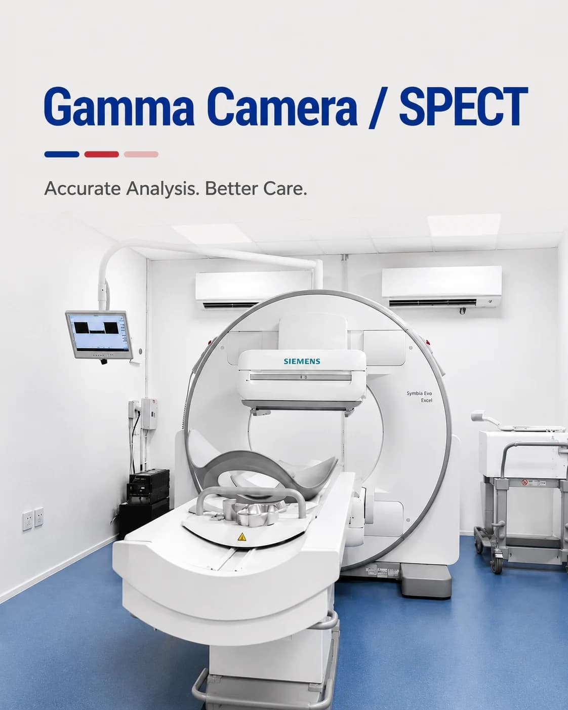
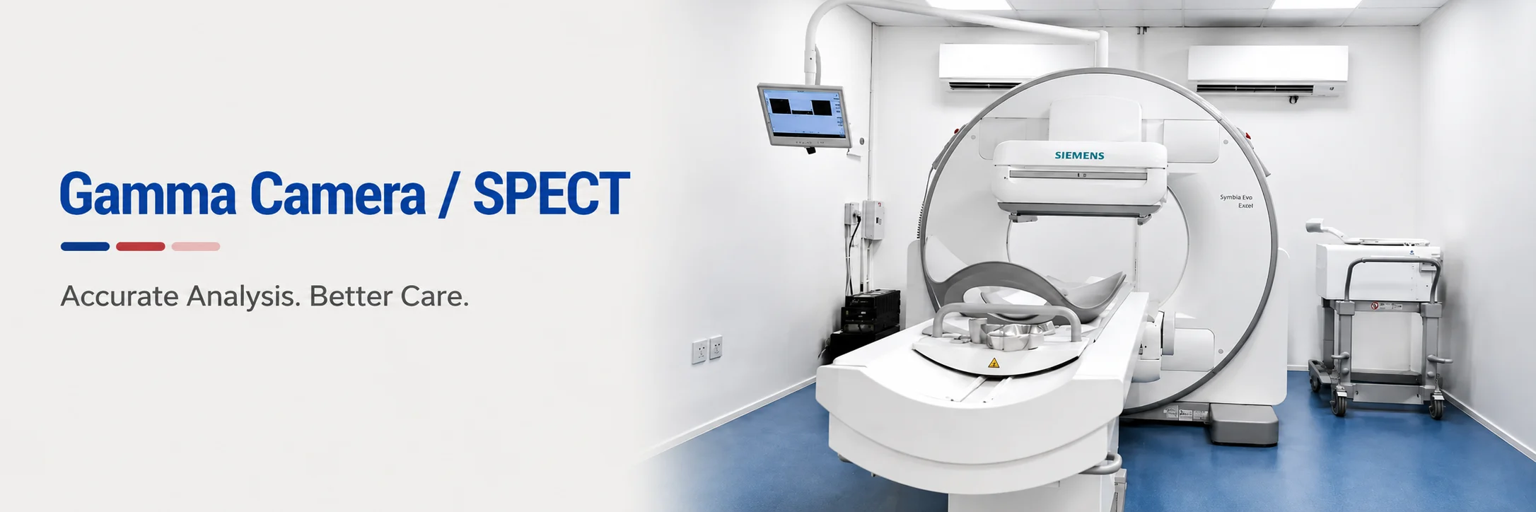
Functional imaging of the heart, bones, kidneys and thyroid using a gamma camera.
A gamma camera detects a small amount of radiotracer given before the scan, building a picture of how an organ is working — its function, not just its form. SPECT adds depth, turning that picture into a three-dimensional study.
At Charak, gamma camera and SPECT studies are used to assess the blood supply to the heart, the activity of bone, the working of the kidneys and the thyroid, each with its own tracer. They answer questions of function that anatomical scans cannot.
What a gamma camera study shows
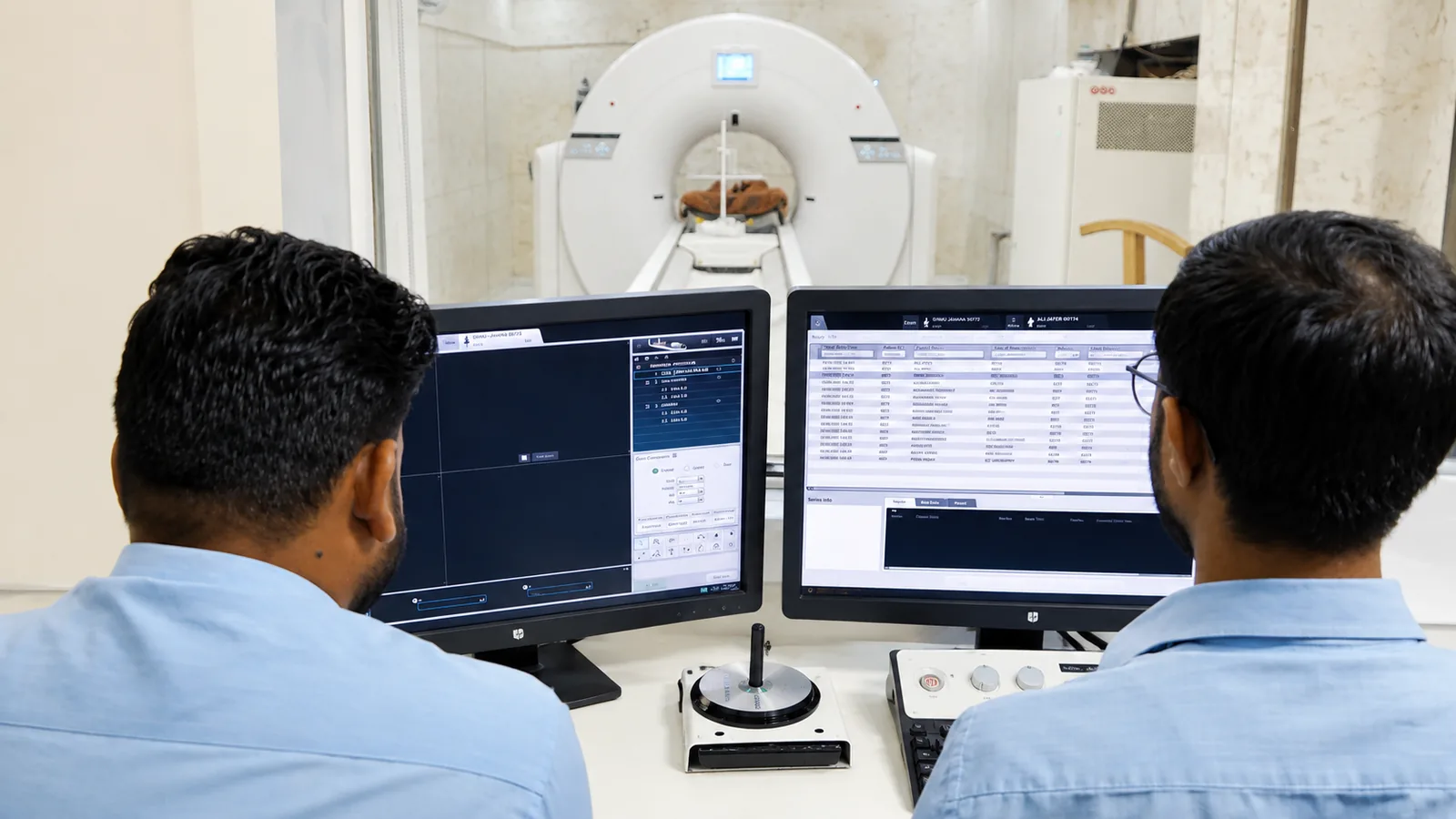
Different tracers are drawn to different organs — one to heart muscle, another to bone, another to the kidneys. The camera maps where the tracer goes and how much arrives, which reveals whether an organ is working normally.
A myocardial perfusion study, for example, shows whether all parts of the heart muscle are getting enough blood, both at rest and under stress — information an ECG or echo alone cannot give.
A gamma camera or SPECT scan may be used for
- Assessing blood supply to the heart muscle (myocardial perfusion)
- Looking for the spread of disease in bone (bone scan)
- Measuring how well each kidney works (renal scan)
- Assessing thyroid function and nodules
- Localising infection or specific functional problems
How to prepare
Preparation depends on which study you are having — a cardiac scan differs from a bone or renal scan. We give you the exact instructions for your specific test when you book.
- Cardiac (perfusion) studies may involve a stress component and specific medication and caffeine rules beforehand
- Bone scans usually need you to drink plenty of water and wait a few hours between the tracer and the scan
- Renal and thyroid studies have their own timing — we explain yours clearly
- Tell us if you are, or might be, pregnant, or are breastfeeding
What to expect on the day
- 01
Tracer injection
The tracer appropriate to your organ is given, usually through a vein in the arm.
- 02
The waiting period
Depending on the study, imaging follows immediately or after a set delay while the tracer reaches the organ.
- 03
Imaging
You lie still while the gamma camera moves slowly around or above you; SPECT studies rotate to build a 3-D view.
- 04
Afterwards
You can resume normal activity; drinking water helps clear the tracer, which fades over the day.
Common questions
How is a gamma camera scan different from a CT or MRI?
CT and MRI show anatomy — the shape and structure of organs. A gamma camera shows function — how well an organ is actually working — using a tracer that the organ takes up. They answer different questions and are often complementary.
Is the tracer safe?
Yes. The amount of radiotracer is very small and short-lived, chosen to give a clear image with the lowest reasonable dose. It clears from the body over hours, and drinking water helps.
Will the scan take a long time?
It varies by study. Some imaging is done shortly after the injection; others need a delay of a few hours between the tracer and the scan. We tell you the timing for your specific test in advance.
From the health library
Plain-language guides written by the team here — what the test measures, how to prepare, and what the result means.
Related services
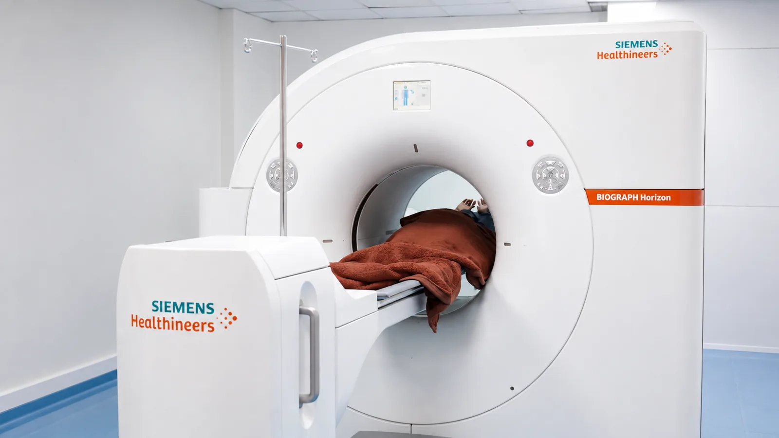
PET-CT Scan
Whole-body imaging that shows how tissue is functioning, not just how it looks.
Read more →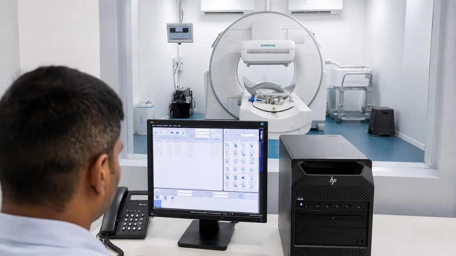
Thyroid Scan
A functional look at the thyroid gland — how active it is, and how it works.
Read more →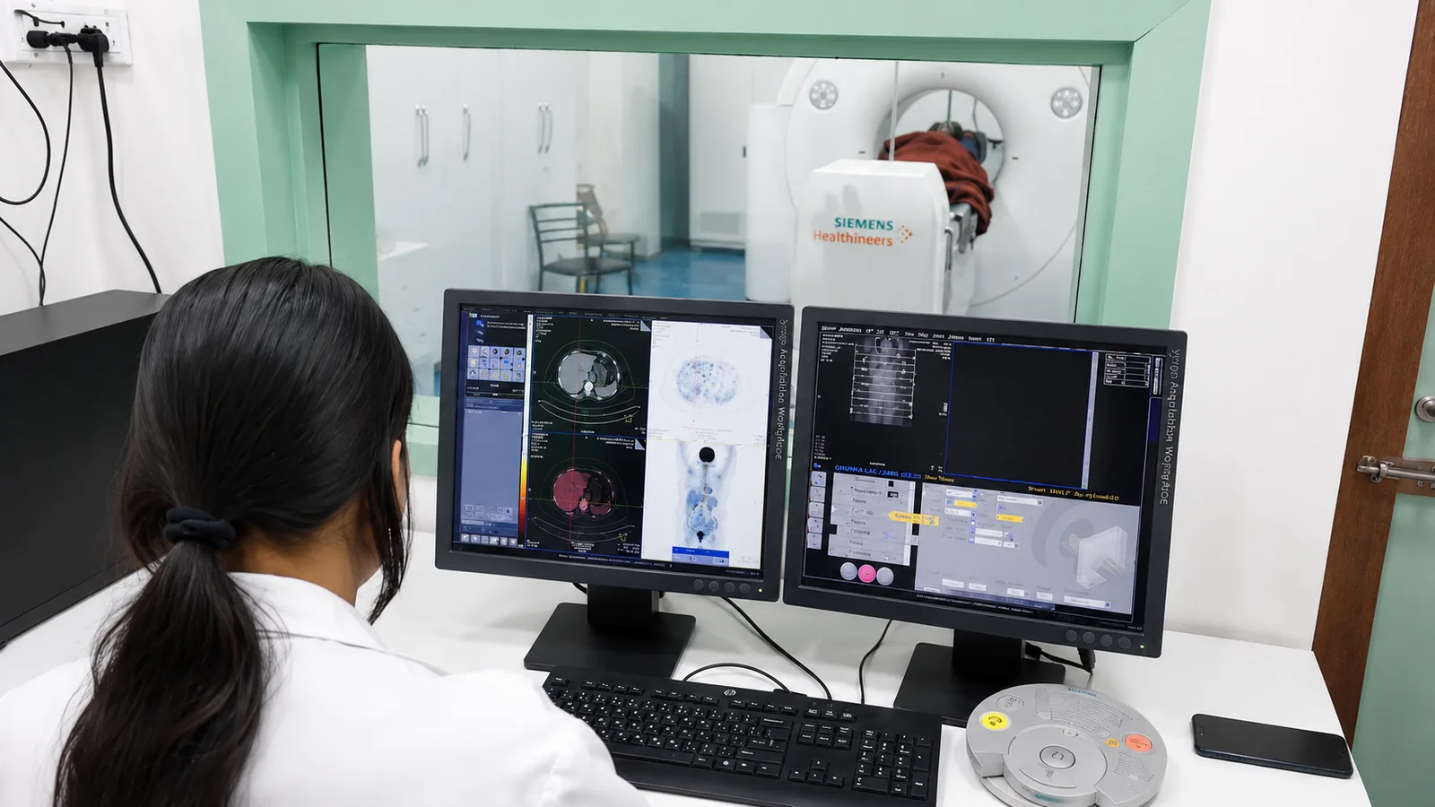
Renal Scan (DTPA / DMSA)
Measuring how well each kidney drains and works — one side against the other.
Read more →