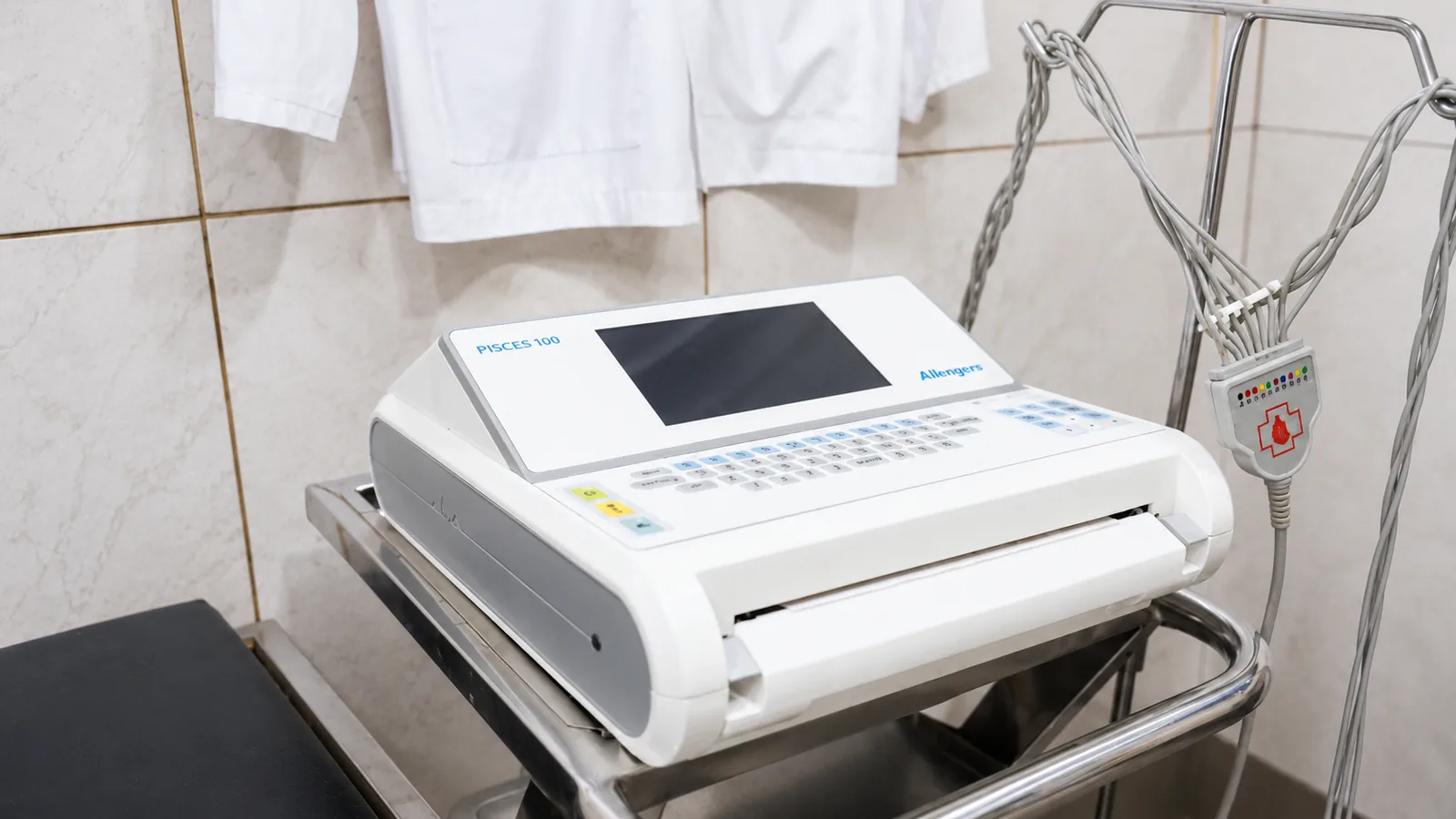
2D Echocardiography in Lucknow
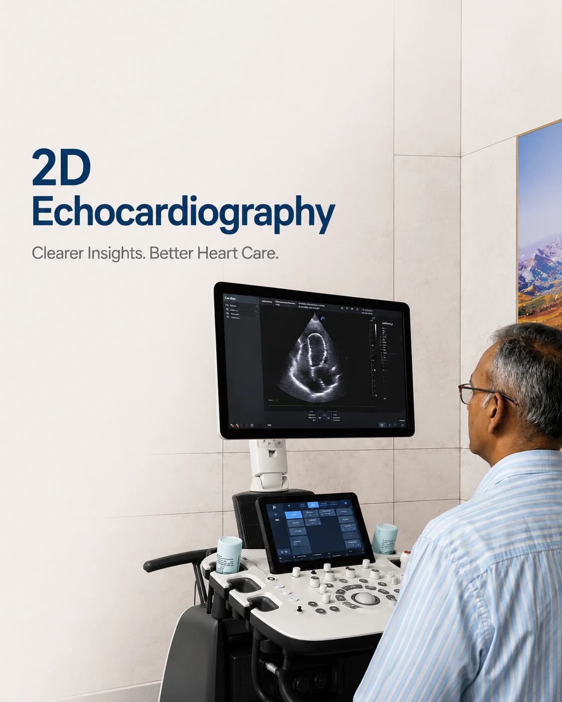
A live ultrasound of the heart — its chambers, valves and pumping strength.
A 2D echocardiogram is an ultrasound of the heart. It shows the heart beating in real time — the size of its chambers, the movement of its valves, and how strongly it is pumping — all without radiation and without any discomfort.
At Charak, echocardiography is performed by trained operators and reported by cardiologists, with colour Doppler added to show how blood flows through the valves. It is the standard test for assessing heart function and is safe to repeat as often as needed.
What an echo measures
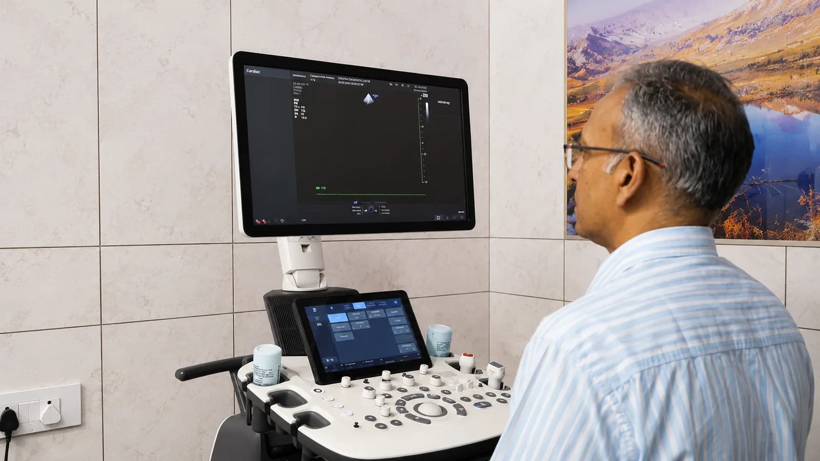
The echo shows the heart's pumping strength as the ejection fraction — a key number in heart failure — and reveals whether the valves are narrowed or leaking. It can also show fluid around the heart and the effect of long-standing high blood pressure on the muscle.
Colour Doppler overlays the flow of blood in colour, so a leaking or narrowed valve is seen as well as measured. Together they give a complete, moving picture of how the heart is working.
An echo may be recommended for
- Breathlessness or suspected heart failure
- A heart murmur or suspected valve problem
- After a heart attack, to assess the pumping strength
- High blood pressure and its effect on the heart
- Palpitations, or before certain treatments and surgery
How to prepare
A standard (transthoracic) echo needs no preparation and no fasting — it is done from the surface of the chest.
- No fasting is required for a routine echo
- Wear a two-piece outfit, as the upper body is exposed for the scan
- Take your medicines as usual
- Bring any previous echo or cardiac reports for comparison
What to expect
- 01
Settling in
You lie on your left side on the couch, and a little gel is applied to the chest for the probe.
- 02
The scan
The operator moves the probe over the chest, capturing the heart from several angles; you may be asked to breathe in and hold.
- 03
Doppler flow
Colour Doppler is used to study the valves; you may hear the rhythmic sound of blood flow, which is normal.
- 04
Afterwards
The gel is wiped off and you can leave straight away; the cardiologist's report follows.
Common questions
Is an echocardiogram the same as an ECG?
No. An ECG records the heart's electrical activity as a trace; an echo is an ultrasound that shows the heart moving — its chambers, valves and pumping strength. They are complementary, and doctors often use both together.
Does an echo involve radiation?
No. Echocardiography uses ultrasound — sound waves — so there is no radiation. It is completely safe and can be repeated as often as your care needs.
What is ejection fraction?
It is the percentage of blood the heart pumps out with each beat — a key measure of pumping strength. A normal value is a reassuring sign; a low one is important in diagnosing and following heart failure.
From the health library
Plain-language guides written by the team here — what the test measures, how to prepare, and what the result means.