Health Library / Women's health
Mammography in Lucknow: when to start, and what the discomfort is for
· 5 min read · Lucknow
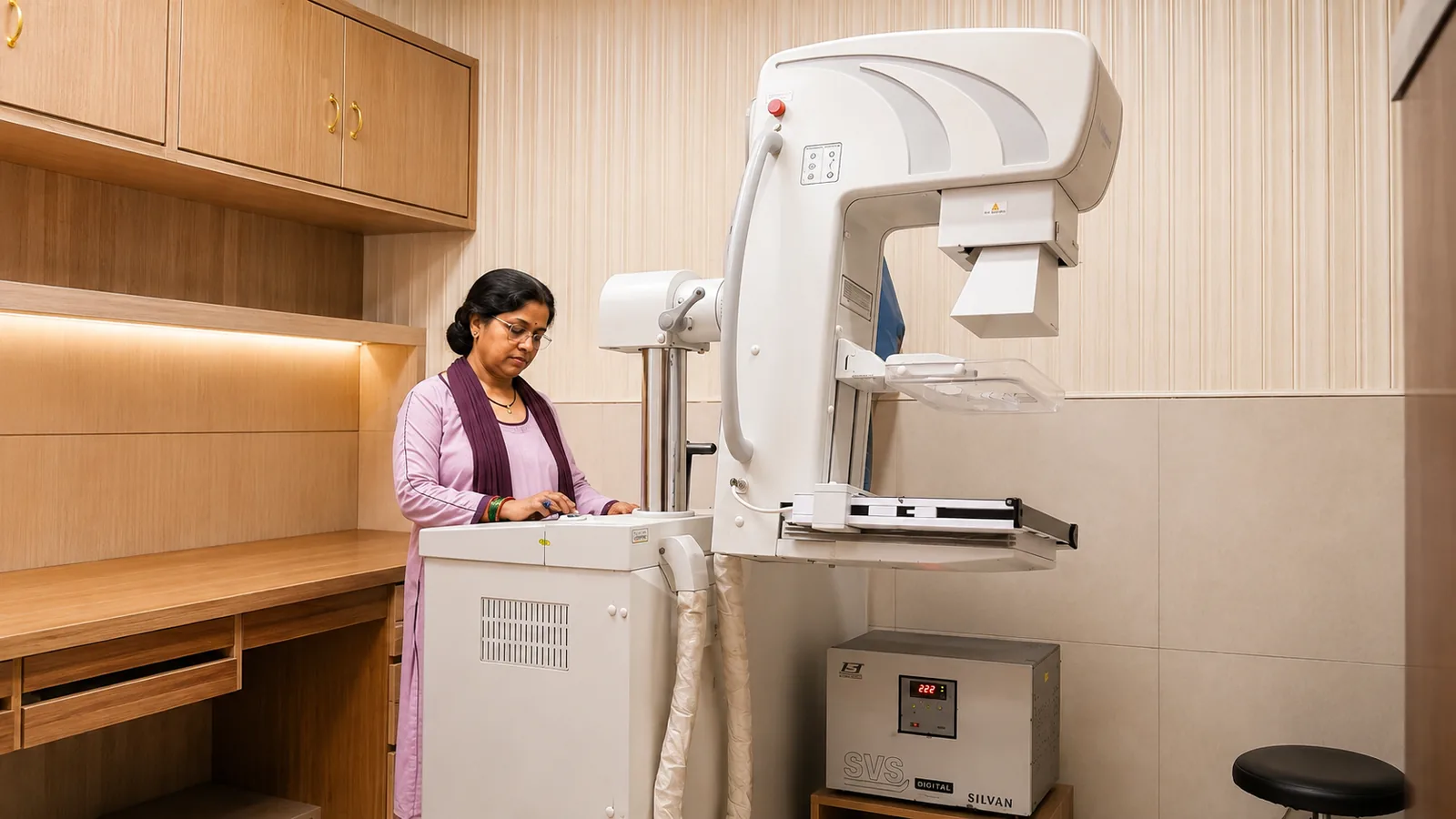
A mammogram is a low-dose X-ray of the breast, and it can find change years before anything can be felt. Breast cancer in India is often diagnosed late, and lateness — not the disease alone — is a large part of why outcomes are worse here than they need to be.
It is also the test most often postponed, usually because of what people have heard about the compression. That part is true, brief, and worth understanding rather than avoiding.
Screening versus diagnostic, and when to start
A screening mammogram is done when there are no symptoms, to look for early change. A diagnostic mammogram is done when there is something specific — a lump, pain, skin or nipple change — and takes additional focused views.
The age to begin screening depends on your risk and on your doctor's advice, and it starts earlier where there is a strong family history or a known genetic risk. Rather than a single rule, treat it as a conversation to have at your next appointment — particularly if a close relative has had breast or ovarian cancer.
- A new lump or thickening in the breast or armpit
- Change in the skin — dimpling, puckering or redness
- Nipple change, inversion, or unusual discharge
- Persistent, localised breast pain
- Any change after previous breast treatment
Why compression, and how to make it easier
The breast is compressed for a few seconds per view. It spreads the tissue so overlapping structures do not hide a small change, holds it still so the image is sharp, and lets a lower radiation dose do the job. Skipping it would mean a blurrier image at a higher dose — the discomfort is buying image quality.
You can make it easier. Book for the week after your period if you still menstruate, when breasts are least tender. Skip talcum powder, deodorant and lotion on the day, because they can show up on the image and be mistaken for calcification. Wear a two-piece outfit so only the upper half needs removing.
Dense breasts, and why ultrasound is added
Dense breast tissue is normal and common, particularly in younger women, but it appears white on a mammogram — and so do many of the changes being looked for. That reduces how much a mammogram can see.
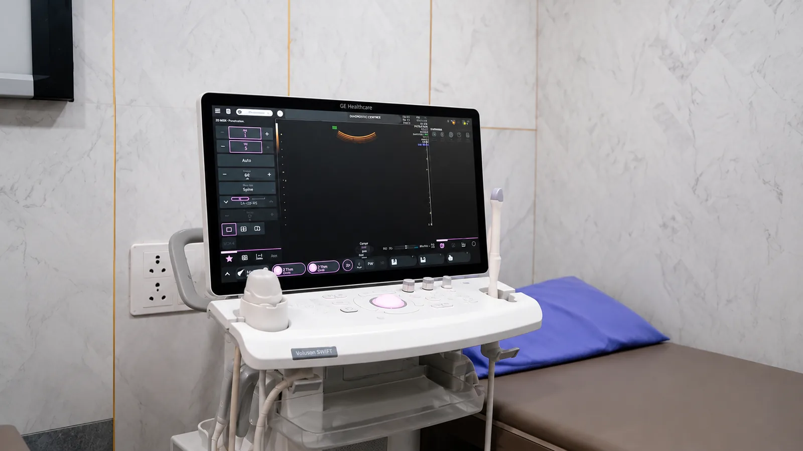
This is why ultrasound is often added rather than substituted: the two together see more than either alone. If you have been told your breasts are dense, ask your doctor whether an ultrasound alongside is appropriate for you.
At Charak, mammography and ultrasound are in the same building at Chowk, examinations are done in private rooms, and reporting is by consultant radiologists. Bring any previous mammograms — change over time is often the finding itself.
This article is general health information, not a diagnosis or a treatment plan. Test results are read alongside your symptoms and history — please discuss yours with your doctor. See our disclaimer.