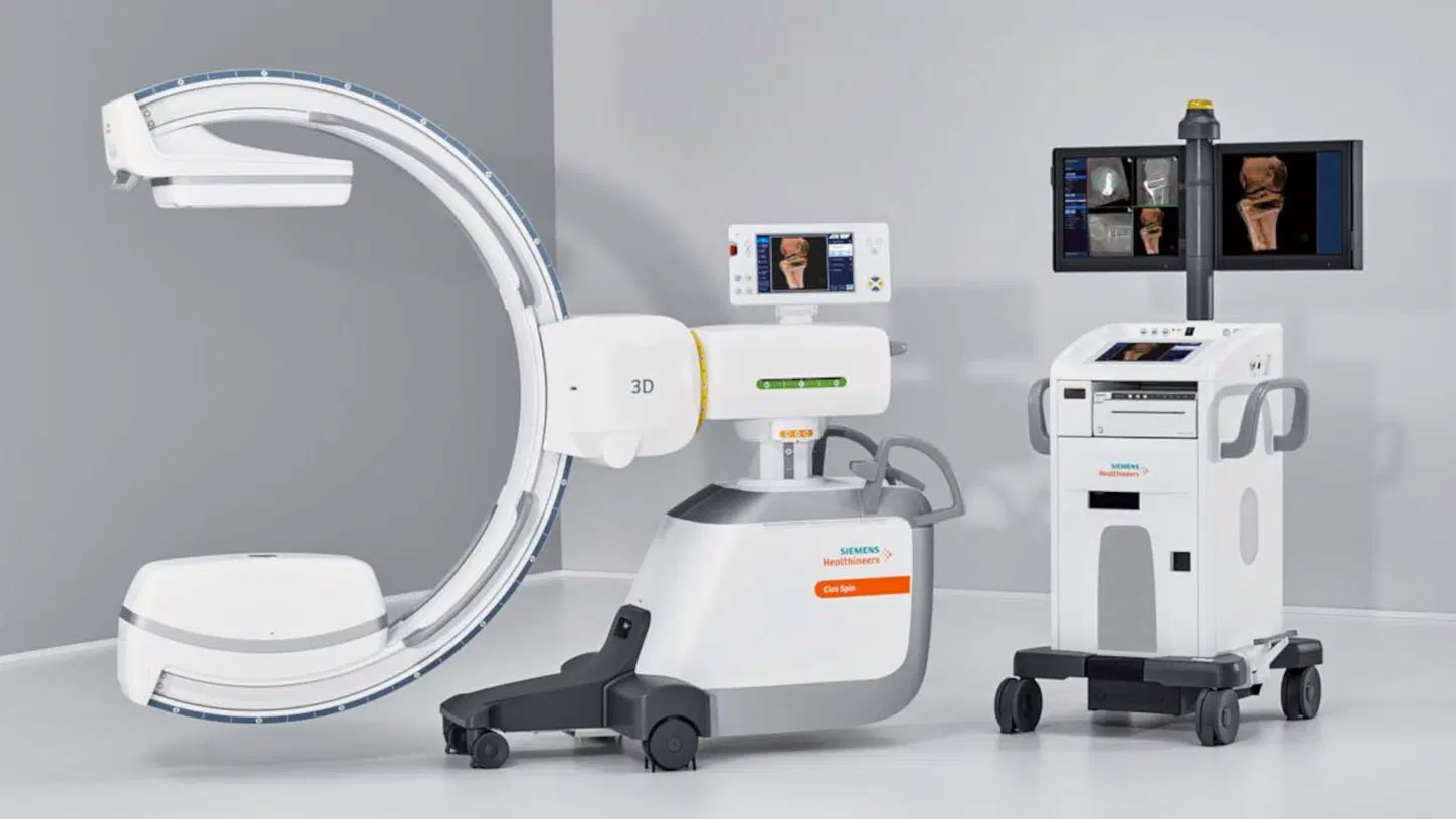
Hysterosalpingography (HSG) is a radiologic procedure used to evaluate the uterine cavity and fallopian tubes in women. It is typically performed by a radiologist or a specialized technician in a radiology department or clinic.
Here's how the procedure works:
-
Preparation: Before the procedure, the patient is asked about their medical history, including any allergies, and a pregnancy test may be performed to ensure the patient is not pregnant.
-
Positioning: The patient is asked to lie on an examination table in a radiology suite. A speculum is inserted into the vagina to visualize the cervix.
-
Catheter Insertion: A thin, flexible tube called a catheter is inserted into the cervix and advanced into the uterine cavity. This allows contrast material (a dye) to be injected directly into the uterus.
-
Contrast Injection: A radiopaque contrast material is injected through the catheter into the uterus. The contrast material is visible on X-ray images and allows for the visualization of the uterine cavity and fallopian tubes.
-
X-ray Imaging: As the contrast material is injected, a series of X-ray images are taken in real-time. These images help to identify any abnormalities in the shape of the uterus, such as uterine fibroids, polyps, or other structural issues.
-
Evaluation of Fallopian Tubes: The contrast material should flow through the uterus and into the fallopian tubes. If there are any blockages or abnormalities in the fallopian tubes, they can be detected during the procedure.
-
Conclusion: Once the necessary images have been captured, the catheter is removed, and the procedure is complete.
HSG is often used to investigate the cause of female infertility, as it can reveal issues like blocked fallopian tubes or structural abnormalities in the uterus. It can also be used to check for problems like recurrent miscarriages or abnormal vaginal bleeding. The results of the HSG can help guide further treatment or fertility interventions.

